Yigong Shi’s Group publishes an article in Cell, reporting the cryo-EM structure of the post-catalytic spliceosome from Saccharomyces cerevisiae
On November 17th 2017, Professor Yigong Shi’s group in the School of Life Sciences, Tsinghua University, published an article entitled “Structure of the Post-catalytic Spliceosome from Saccharomyces cerevisiae” in the prestigious research journal Cell. The paper reported the cryo-EM structure of post-catalytic spliceosome, a crucial complex for RNA splicing in Saccharomyces cerevisiae, at the average resolution of 3.6 angstrom. This structure illustrates the mechanism of spliceosome remodeling after RNA splicing reaction and 3’-splice site (3’SS) recognition. This high resolution structure reveals unexpected features of the 3'SS recognition during the second-step RNA splicing reaction, the transesterification occurred in the second step and the release of the mature mRNA.
In 1977, scientists found that adenovirus mRNA could not form a continuous double-stranded RNA-DNA hybridization duplex with its corresponding DNA transcription template for the first time. Instead, single-stranded DNA bulges were extended at different positions in the hybridized double-stranded RNA-DNA duplex. This important result showed that the transfer of genetic information from DNA to mRNA not only involved transcription but also pre-mRNA splicing, which is to further remove "useless" genetic information and connect useful genetic information. "Useless" genetic information, which is called the intron, cannot be translated, and the useful genetic information which can be translated by ribosome is called the exon. RNA splicing widely exists in eukaryotes, the average number of introns per gene increases as one looks from simple single-celled eukaryotes, such as yeast, through higher organisms such as worms and flies, all the way up to humans. Some pre-mRNAs can be spliced in more than one way. Thus, mRNAs containing different selections of exons can be generated from a given pre-mRNA. Called alternative splicing, this strategy enables a gene to give rise to more than one polypeptide product.
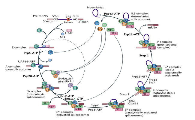
Splicing of pre-messenger RNA (pre-mRNA) is an important and complicated step of central dogma in eukaryotic cells. This process is accomplished by a dynamic mega-complex known as the spliceosome. From the discovery of RNA splicing in 1977 to the beginning of this century, scientists initially established the process of assembly and disassembly of the spliceosome by immunoprecipitation, gene knockout, cross-linked mass spectrometry, and in vitro splicing reaction and so on. During an RNA splicing reaction, an intron is removed through two successive transesterification reactions in which phosphodiester linkages within the pre-mRNA are broken and new ones are formed. To catalyze splicing of the pre-mRNA, spliceosome undergoes extremely precise stepwise assembly, subsequently forming a series of spliceosomal complexes. According to the reaction state and biochemical characterization, these complexes can be defined as tri-snRNP, B, Bact , B*, C, C*, P, and ILS complexes (Figure 1).

Figure 1. A cartoon diagram of RNA splicing.
(Shi Y. Nature Reviews Molecular Cell Biology, 2017.)
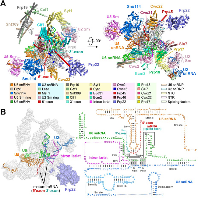
Post-catalytic spliceosome (P complex) is a highly dynamic and transient state and is difficult to purify under normal physical conditions. In their recent Cell paper, Shi and colleagues enriched P complex samples in S.cerevisiae cells through over-expression of an ATPase-defective Prp22 mutant, leading to the inhibition of mature mRNA release in vivo. Then they acquired a good P complexes sample by applying TAP purification strategy and determined the three-dimensional structure of post-catalytic spliceosome (P complex) at the overall resolution of 3.6 angstrom by single particle cryo-EM, and built the atomic model (Figure 2). This structure displays the overall structure of the spliceosome and the organization of spliceosomal protein and RNA components after the two-step transesterification reactions during RNA splicing. In this structure, we can clearly see the two exons, which is separated by an intron before splicing, is covalently linked, forming a mature mRNA and anchored in spliceosomal active site through base-paring with U5 snRNA. Remarkably, this structure first reveals the recognition mechanism of 3’SS of the pre-mRNA. The dinucleotide AG at the 3’SS is separately base-paired with adenine nucleotide in BPS and the first guanine nucleotide at 5’SS, and further stabilized through stacking interaction by G of 5’SS and U6 snRNA. Therefore, this structure provides important structural evidence to illustrate the mechanism of 3’SS recognition and mRNA release by Prp22, promoting and advancing our understanding of pre-mRNA splicing.

Figure 2. Structure of the Post-catalytic Spliceosome, Known as the P Complex, from S. cerevisiae.
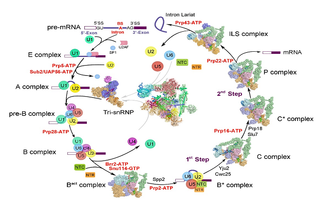
Shi's group has been working on capturing the high-resolution structures of the spliceosome at different reaction states to explore the molecular mechanism of RNA splicing. Since August, 2015, Shi’s group have reported seven high resolution structures of the crucial spliceosomal complexes, that is, the S.pombe intron lariat spliceosome ILS complex at 3.6 angstrom, the pre-assembled U4/U6.U5 tri-snRNP at 3.8 angstrom, the activated spliceosome Bact complex at 3.5 angstrom, the catalytic step I spliceosome C complex at 3.4 angstrom, the step II catalytically activated spliceosome C* complex at 4.0 angstrom, the ILS complex at 3.5 angstrom and the newest published post-catalytic spliceosomal P complex at 3.6 angstrom from S.cerevisiae. These seven high resolution structures almost cover all the working states of spliceosome assembly, catalysis and disassembly, providing extremely abundant information which subsequently aids the development of the RNA splicing research. Prof. Yigong Shi has just won the 2017 Future Science Prize (Life Science Award) for his elucidation of high-resolution structures of the eukaryotic spliceosome, revealing the active-site and the molecular-level mechanism of this key complex in mRNA maturation.

Figure 3. Cryo-EM structure of yeast spliceosome solved by Shi lab.
Prof. Yigong Shi is the corresponding author. Rui Bai (PhD student from the School of Life Sciences), Dr. Chuangye Yan (post-doc fellow from the School of Life Sciences and Center for Life Sciences), and Ruixue Wan (PhD student from the School of Medicine) are the co-first authors of this paper. Dr Jianlin Lei provided technical support on the EM data collection. EM images were acquired at the Tsinghua University Branch of the China National Center for Protein Sciences (Beijing). Data processing was performed on the “Explorer 100” cluster system of Tsinghua National Laboratory for Information Science and Technology, the Computing Platform of China National Center for Protein Sciences (Beijing). This research was funded by the Beijing Innovation Center for Structural Biology and the National Natural Science Foundation of China.
The original link:
http://www.cell.com/cell/fulltext/S0092-8674(17)31264-3
Related publications:
http://science.sciencemag.org/content/early/2016/01/06/science.aad6466
http://science.sciencemag.org/content/early/2015/08/19/science.aac8159
http://science.sciencemag.org/content/early/2015/08/19/science.aac7629
http://science.sciencemag.org/content/early/2016/07/20/science.aag0291
http://science.sciencemag.org/content/early/2016/07/20/science.aag2235
http://science.sciencemag.org/content/early/2016/12/14/science.aak9979.full
http://www.cell.com/cell/fulltext/S0092-8674(17)30954-6
(Edited by Guo Lili, Zhu Lvhe)

